Contacts: Derek Wright or Stephen Kolomyjec
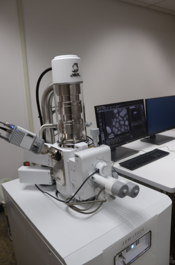
The IT-200 is a variable pressure tungsten filament SEM and is capable of resolving features as small as 3nm. Available detectors include: SE, BSE, EDS (elemental analysis), and a STEM converter for transmission imaging. The EDS detector allows both surface spot analysis of elemental composition and element mapping. JEOL PA3 software for automated particle imaging and analysis is also available.
Specifications:
Resolution: 3nm (30KeV, SE, High Vacuum)
EDS (30mm2 detector): ~1-3 µm resolution, Elements: Boron-Uranium. Detection limits ~0.1-1.0 wt% (better sensitivity for light elements)
Magnification: Up to 300,000x
Sample Requirements:
Vacuum Stable (dry, non-volatile)
Dimensions less than a few cm, most samples fit on a 32 mm (1 1/4 inch) or smaller holder
Samples for STEM Imaging must fit on a 3mm TEM Grid and should be <100nm thick
Accurate quantitative EDS analysis requires samples to be flat & polished
Nonconductive samples must be gold/carbon coated or imaged under low vacuum conditions
Contacts: Derek Wright or Mark Zierden
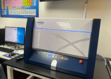
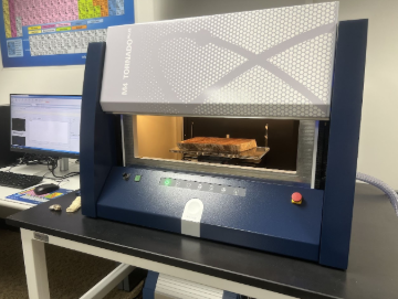
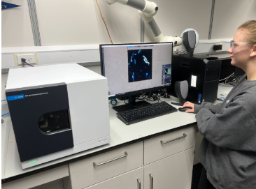
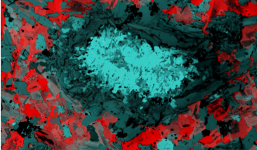
The Bruker M4 Tornado Plus micro X-Ray Fluorescence spectrometer (µXRF) is a state of the art instrument capable of both spot analysis and imaging of the elemental distributions of large specimens (16×19 cm) at high spatial resolution (~20 µm). For biological specimens, the penetration of the x-ray beam is sufficient to permit analysis of internal element distributions, making this instrument uniquely suited to studies of element uptake, bioaccumulation, and homeostasis.
Specifications:
EDS (twin 60mm2 detectors with light element windows): ~20 µm resolution (Rh source with polycapillary optics), Elements: Carbon-Uranium. Detection limits ~0.001-1.0 wt% (better sensitivity for heavy elements)
Aperture Management System for increased depth of field
Second source: W Source with collimator optics (0.5mm-4.5mm)
He or vacuum for light element analysis
X-ray penetration allows for imaging of the internal element distribution of specimens with a light element matrix such as biological tissues
Sample Requirements:
Sample dimensions up to 16×19 cm wide (maximum imaging area) and several cm thick. Sample topography should be less than 1cm difference for optimal imaging. Note: larger specimens can be imaged as long as they fit inside the chamber and weigh <7kg (15lbs.)
Liquid, wet, or oily samples can be imaged in air or He atmosphere
Accuracy of quantitative EDS analysis is improved if samples are flat & polished
Contacts: Derek Wright or Benjamin Southwell
The Agilent 8700 LDIR in an infrared chemical imaging system that is capable of point analysis, automated particle analysis, and chemical distribution mapping. As a molecular spectroscopy technique, LDIR responds to variations in molecular or mineral composition. Unlike conventional FTIR microscopes, LDIR uses a rapid scanning tunable quantum cascade laser to collect infrared data between 975-1800cm-1.
Specifications:
Resolution: ~5 µm
Imaging Modes: Reflectance, Transflectance using IR reflective slides
Sample Requirements:
Dry
Maximum Dimensions 25x75mm (standard microscope slide, also thin section slides can be accommodated)
Flat (geologic specimens benefit from polishing)
Thickness/particle size <500 µm for transflelctance mode
Specimens <1cm thick for reflectance mode
Contacts: Derek Wright or Stephen Kolomyjec
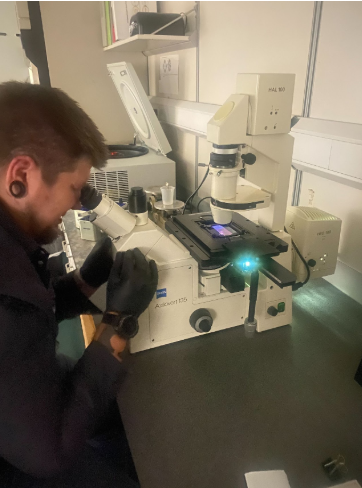
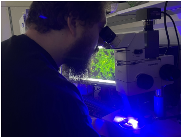
Fluorescence microscopy is one of the most widely used techniques to localize structures or compounds of interest within cells and tissues, and has utility for fluorescent inorganic materials as well. We have three research grade widefield fluorescence microscopes available for general use:
Zeiss Axioskop 2 Compound (CRW 265, Optical Microscopy Lab)
Filters: DAPI, FITC, TRITC
Contrast Modes: Transmitted Brightfield, Darkfield, Phase Contrast
Objectives: Plan Neofluar 5x (ach) 10x (0.3NA), 20x (0.5NA) 40x (0.75NA), and 100x (1.3NA,Oil)
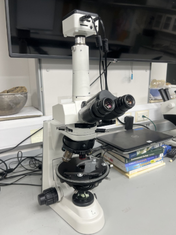
Nikon Eclipse E600 Compound (CRW 265, Optical Microscopy Lab)
Filters: DAPI, FITC, TRITC
Contrast Modes: Transmitted Brightfield, Darkfield, Phase Contrast, Nomarski-DIC
Objectives: Plan Neofluar 4x(0.13NA), 10x (0.3NA), 20x (0.5NA), 40x (0.75NA), and 100x (1.3NA,Oil)
Zeiss Axiovert 135 Inverted (CRW 231)
Filters: DAPI, FITC, TRITC
Contrast Modes: Transmitted Brightfield, Darkfield, Phase Contrast, Nomarski-DIC
Objectives: Plan Neofluar 10x(0.3NA,Ph), 20x (0.4NA,Ph), 20x (0.5NA,DIC) 40x (0.75NA,DIC), and 100x (1.3NA,DIC, Oil)
Contact: Paul Kelso
Polarized lightmicroscopy is a technique used to identify minerals, fibers, and certain biologic structures.
Nikon Eclipse 50iPOL Petrographic Microscope: (CRW 341)
Transmitted polarized light
2x, 4x, 10x, 20x, 40x objectives
Bertrand lens for conoscopy
Contacts: Derek Wright and Stephen Kolomyjec
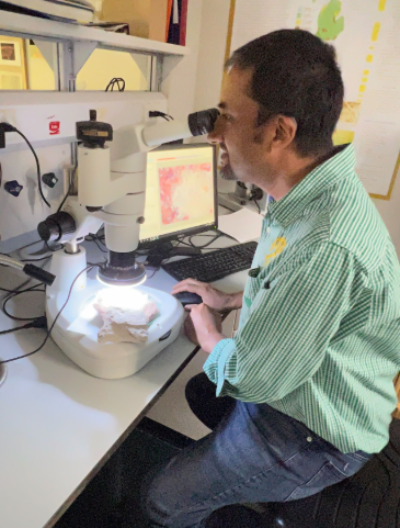
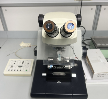
Stereomicroscopes are used to view three dimensional samples under magnification and are especially useful for examining larger, intact samples. We have three research stereomicroscopes available for general use. A Nightsea fluorescence adapter is also available for fluorescence stereomicroscopy.
OlympusSZH and SZH-10 Stereomicroscopes: (CRW 258)
6.5-65x/7-70x magnification
Illumination: Transmitted brightfield and darkfield, Oblique, Ring light/polarizing ring light
Nikon SMZ1000N Stereomicroscope: (CRW 265)
8–80x magnification
Illumination: Transmitted brightfield and darkfield, Oblique, Ring light/polarizing ring light
Contacts: Derek Wright and Stephen Kolomyjec
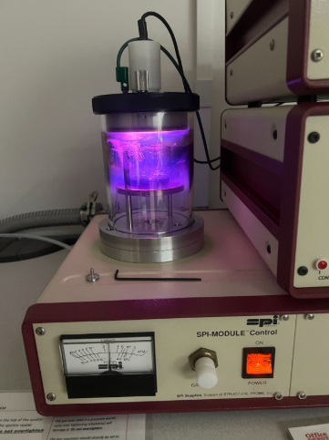
To image and analyze samples in the SEM under high vacuum conditions (highest resolution), the samples must be electrically conductive. Nonconductive specimens are therefore typically coated with an ultrathin (a few nm) layer of either gold (optimal for imaging) or carbon (optimal for EDS analysis). The SPI coater has modules for both sputter coating and carbon coating.
Contacts: Stephen Kolomyjec or Derek Wright
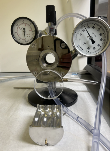
To image and analyze samples in the SEM they must dry. For many hydrated specimens such as soft biological tissues, drying without deforming them can be challenging. The most widely used method for overcoming this challenge is to use supercritical carbon dioxide with a critical point dryer. We can also perform chemical drying using HMDS and lyophilization (freeze drying), which may be suitable for some samples.
Contacts: Barba Evans
Transmission electron imaging (STEM) requires samples to be section to 100nm or less in thickness with an ultramicrotome. The Leica Ultracut R can section at thicknesses of 25-5000 nm.
Contacts: Derek Wright, Hari Kandel, or Paul Kelso
Contact: Paul Kelso
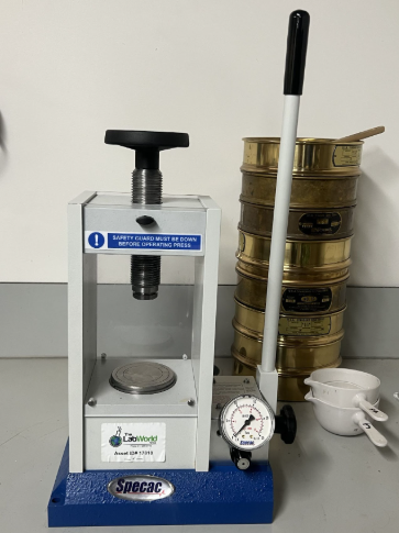
The Geology Program has a fully fitted laboratory for geologic sample preparation, including geologic thin sections. Equipment includes various rock saws, a drill press with 1in. coring bit, lapidary wheels, sieves & shakers, etc.
Additionally, the Engineering Department has a full service machine shop if additional equipment is required. Contact: Ronald Throener, Mechanical Engineering Laboratory Engineer – [email protected], 906-635-2595
Additional instrumentation for biologic & chemical analysis/characterization includes: ICP-MS, MP-AES, LC-MS/MS, LC-TOF-MS, HPLC-PDA, MPLC, GC-MS/MS, Headspace GC-MS, GC-FID/ECD, UV-Vis, FTIR, 400mhz NMR, Fluorescence/Luminescence Spectroscopy, Ion Chromatography, PCR, qPCR, ddPCR, Next Gen Sequencing, Fragment Analysis, and Electrochemistry.